Spider Veins (Telangiectasia): Small, low-flow lesions typically found on the face and legs.Cherry Angiomas: Red, cutaneous hemangiomas appearing on the torso, arms, and legs.
Port-Wine Stains: Capillary malformations that appear on the face, neck, and breast vascular regions.
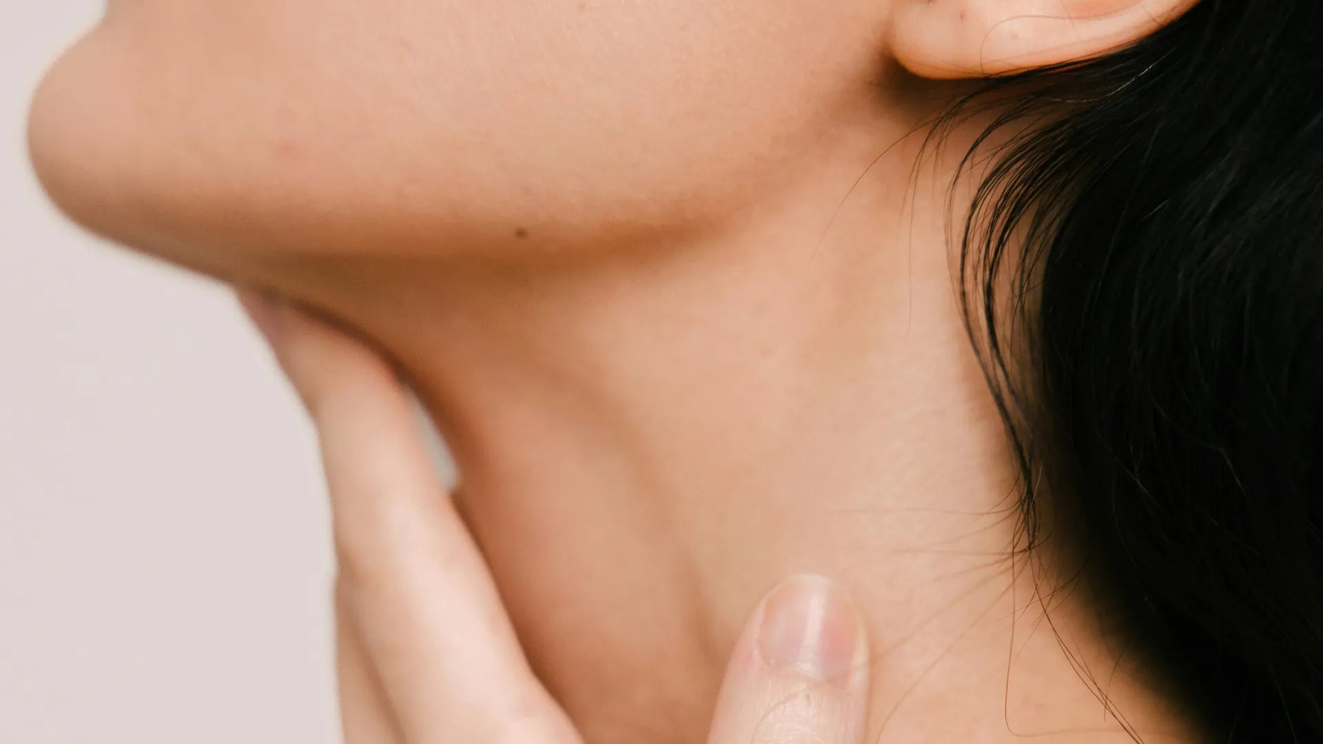
Rosacea-Related Redness: Persistent vascular ectasia mainly on the cheeks and nose.
Venous Lakes: Bluish vascular malformations often seen in sun-exposed areas like the lips and ears.
Infantile Hemangiomas: Benign vascular tumors usually found on the head, neck, and torso at birth.
Arteriovenous Malformations (AVMs): Abnormal arterial-venous connections, which may cause extensive lesions in soft tissues and skin.
Macrocystic & Microcystic Lesions: Found in lymphatic malformations, affecting fluid-fluid levels in the tissues.
Sturge-Weber Syndrome Lesions: Rare vascular malformations associated with the trigeminal nerve.
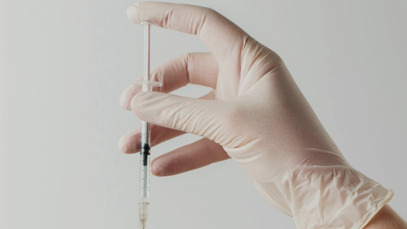
Vascular lesions can present in different forms, ranging from harmless cosmetic concerns to more complex conditions requiring medical evaluation. These lesions develop due to abnormal blood vessel growth or increased blood flow near the skin’s surface. While many are benign, certain characteristics may indicate a need for further assessment.
- Red, purple, or blue patches on the skin.
- Clusters of abnormal blood vessels forming spindle cell hemangiomas.
- Raised or flat soft tissue masses that persist or change in texture.
- Occasional swelling or sensitivity in the affected area.
- Hyperintense masses on imaging, indicating a proliferative phase.
If a vascular lesion grows rapidly, presents heterogeneous enhancement, or has aggressive features, a clinicopathologic evaluation is recommended.